This is a technical note for tracheal intubators for performing tracheal intubation during the COVID-19 pandemic and beyond. Up until July 2, 2021, there have been 182,319,261 confirmed cases and 3,954,324 deaths. Unfortunately, it has been estimated that thousands of healthcare workers have died from the disease. It is not easy to know exactly how many airway managers contracted COVID-19 while fulfilling their vital duties of airway management during the pandemic.
All the relevant principles have been referenced in all available consensus guidelines for airway management in patients with COVID-19. However, the scarcity of medical resources and exhausted capacity in the real world might require alternative strategies at the scene. We need to find an affordable, accessible and available strategy to accomplish the goals of tracheal intubation during the pandemic (e.g., safe, accurate, and smooth).
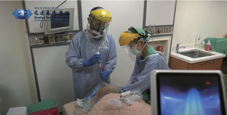
Here, we present our experiences of applying a plastic sheet as an ancillary physical barrier against contagious droplets and secretions from the patient’s airway in Taiwan.1,2 Meanwhile, we use a video-assisted intubating stylet technique to perform tracheal intubation. A brief description of how to prepare such a plastic sheet is shown in Figure 1, and how to apply it with various intubating tools is shown in Figure 2.
Download full-size image
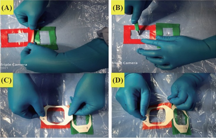
(A) Mark the areas. (B) Cut the crosses with a surgical blade. (C) Tape adhesive films over the cross. (D) Puncture a hole at the center of the cross with a needle.
Download full-size image
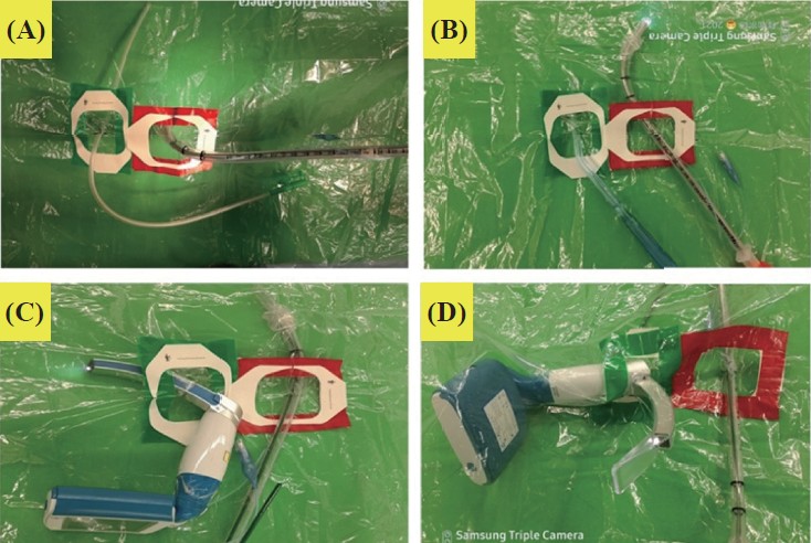
(A, B) One hole for passing the intubating stylet and the other hole for the suction tube. (C, D) One hole for introducing video laryngoscope and the other hole for an endotracheal tube. Notice that one should insert the camera module first and then mount the disposable blade onto the module from the inner side of the sheet.
First, we prepared a transparent and soft plastic sheet (e.g., excised from a plastic trash bag made of ethylene vinyl acetate, 0.05–0.10 mm in thickness; 50 × 80 cm2 in size). We marked two small areas on the plastic sheet (Figure 1A) and cut a small cross with a surgical blade in the center of each marked area (Figure 1B). Then, we covered the marked area with a small transparent adhesive film dressing (e.g., TegadermTM, Figure 1C). Finally, we used a large bore needle to make a small nick at the center of the cross on each film (Figure 1D). Then, it was ready for applying intubating tools, shown in Figure 2.
If the intubating stylet technique was preferred (Figure 2A and 2B), one hole was used for passage of the stylet, and the other hole was for various suction tubes. On the other hand, if video laryngoscopy was the preference, one hole was used to introduce the laryngoscope, and the other hole was for passing the endotracheal tube (Figure 2C ad 2D). During the laryngoscope setup, in order not to damage the adhesive film, which functions to minimize the defect in the plastic sheet and to prevent contamination from the patient, we recommend the following technique. First, the laryngoscope camera module is inserted through the hole (Figure 2C). Then, the disposable blade is mounted over the module from the inner side of the sheet (Figure 2D). In this way, the adhesive TegadermTM film is kept intact.
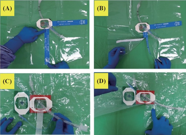
After the endotracheal tube is introduced into the trachea, the plastic sheet is carefully removed in order to prevent further contamination. With this goal in mind, we tear the plastic sheet along the readymade seam line (Figure 3A), until reaching the perimeter of the hole for entry of the endotracheal tube (Figure 3B). In this way, we are able to remove the plastic sheet while leaving the endotracheal tube in a secured position. For a home-made plastic sheet, we first cut the sheet along a straight line to the perimeter covered by TegadermTM and reseal the cut edges with tape (Figure 3C). Later, we can split apart the sheet by simply peeling off the tape (Figure 3D).
Download full-size image
(A) A commercial plastic sheet product with two ready-made seam lines. (B) The plastic sheet can easily be torn apart along each seam line to the perimeter of the area covered by TegadermTM. (C) When using a homemade plastic sheet, one can cut a straight line to the TegadermTM-covered perimeter and reseal the cut edges with tape. (D) One can split apart the sheet by removing the tape from the pre-cut line.
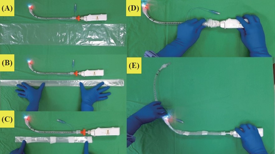
After endotracheal intubation, a valid safety concern remains for the dirty stylet tip. The intubator should always remove and secure the stylet with great care, but we also suggest another practical strategy to help with infection control. We use an umbrella plastic sleeve (Figure 4A), folded to a proper width (Figure 4B), and tailored to fit the stylet (Figure 4C). We then fold the sleeve like an accordion, mounting it onto the proximal end of the stylet, and then insert the stylet into the endotracheal tube (Figure 4D). Later, while withdrawing the stylet from the tube, the compressed sleeve will naturally elongate and wrap up the stylet (Figure 4E). To protect the main body part (the liquid-crystal display panel and main power handle) of the video intubating stylet from possible contamination, one can cover it with a transparent plastic zip lock bag or plastic food wrap. The stylet, after being disconnected from the main body part, is then cleaned and disinfected. It should be noted that one should follow the local infection control policies and procedure to clean and disinfect the intubating tools.
Download full-size image
Find an umbrella plastic sleeve (A), fold it to a proper width (B), and tailor it to size for the stylet (C). (D) Accordion fold the sleeve and mount it onto the proximal end of the stylet and then insert the stylet into the endotracheal tube. (E) When withdrawing the stylet from the tube, the folded sleeve will elongate and wrap up the stylet.
Please find the supplementary materials of Supplement Video 1 for a practical demonstration of the procedure and Supplement Video 2 for a technical note.
References
| 1 |
Yang YL, Huang CH, Luk HN, Tsai PB.
Adaptation to the Plastic barrier sheet to facilitate intubation during the COVID-19 pandemic.
Anesth Analg. 2020;131(2):e97-e99.
|
| 2 |
Luk HN, Yang YL, Tsai PB.
Application of plastic sheet
barrier and video intubating stylet to protect tracheal
intubators during coronavirus disease 2019 pandemic:
a Taiwan experience [Erratum in Cell Transplant.
2021:30:9636897211027078].
Cell Transplant. 2021;30:963689720987527.
|
Supplement
Download full-size image
The video is available at http://doi.org/10.6859/aja.202109/PP.0003

Download full-size image
The video is available at http://doi.org/10.6859/aja.202109/PP.0003