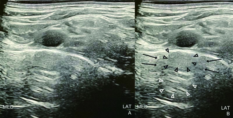
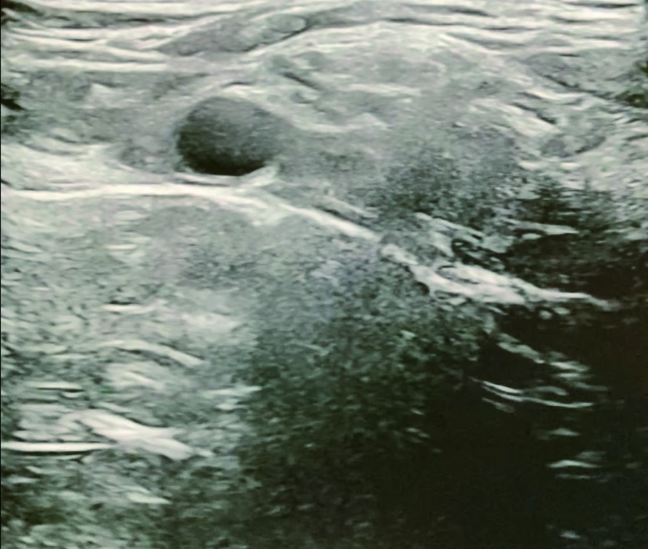
Preliminary scanning for a supraclavicular brachial plexus block showed the brachial plexus trunks lateral to the subclavian artery, above a hyperechoic line elevated from the pleura, suggesting the first rib (Figure 1 and Video 1). However, mirror-image artifacts of the subclavian artery were observed below the supposed first rib, becoming prominent during respirations.
The supraclavicular brachial plexus block is often achieved by guiding the block needle to the "corner pocket" between the subclavian artery and the first rib. 1 The latter provides a backstop to avoid pleural puncture. Misinterpretation of the hyperechoic line as the first rib could have resulted in a pneumothorax.
Mirror image artifacts occur when ultrasound waves encounter a highly reflective surface. Some ultrasound waves bounce off this surface, hit the structure of interest, reflect to the reflective surface again, and finally return to the transducer. Ultrasound machines assume one single round-trip per ultrasound wave, and interpret this longer path as if the structure were located deeper in the body, creating a duplicate image opposite the reflective surface. 2 Mirror image artifacts are not possible through bone due to bone's high attenuation of ultrasound energy and the high acoustic impedance mismatch between soft tissue and bone. Additional reverberation artifacts of the subclavian artery are seen deeper in the lung instead of the acoustic shadowing expected below a rib.
This patient underwent first rib resection for thoracic outlet syndrome and thus had the sonographic view described above. Supraclavicular brachial plexus sonography may look straightforward, but recognition of common artifacts can prevent inadvertent catastrophe.
Download full-size image
Panel A shows an unannotated view. Panel B marks the mirror image artifact (black arrowheads), secondary reverberation artifact (white arrowheads), and acoustic shadowing flanking these artifacts (black arrows) deep to the subclavian artery. Abbreviations: LAT, lateral; MED, medial.
Download full-size image
Note the synchronized movement of all artifacts with the subclavian artery, and the smooth, continuous contour of the pleura. The video can be accessed at https://doi.org/10.6859/aja.202603/PP.0001.
Funding
Support was provided solely from institutional and/or departmental sources.
Conflict of Interest
The authors declare no competing interests.