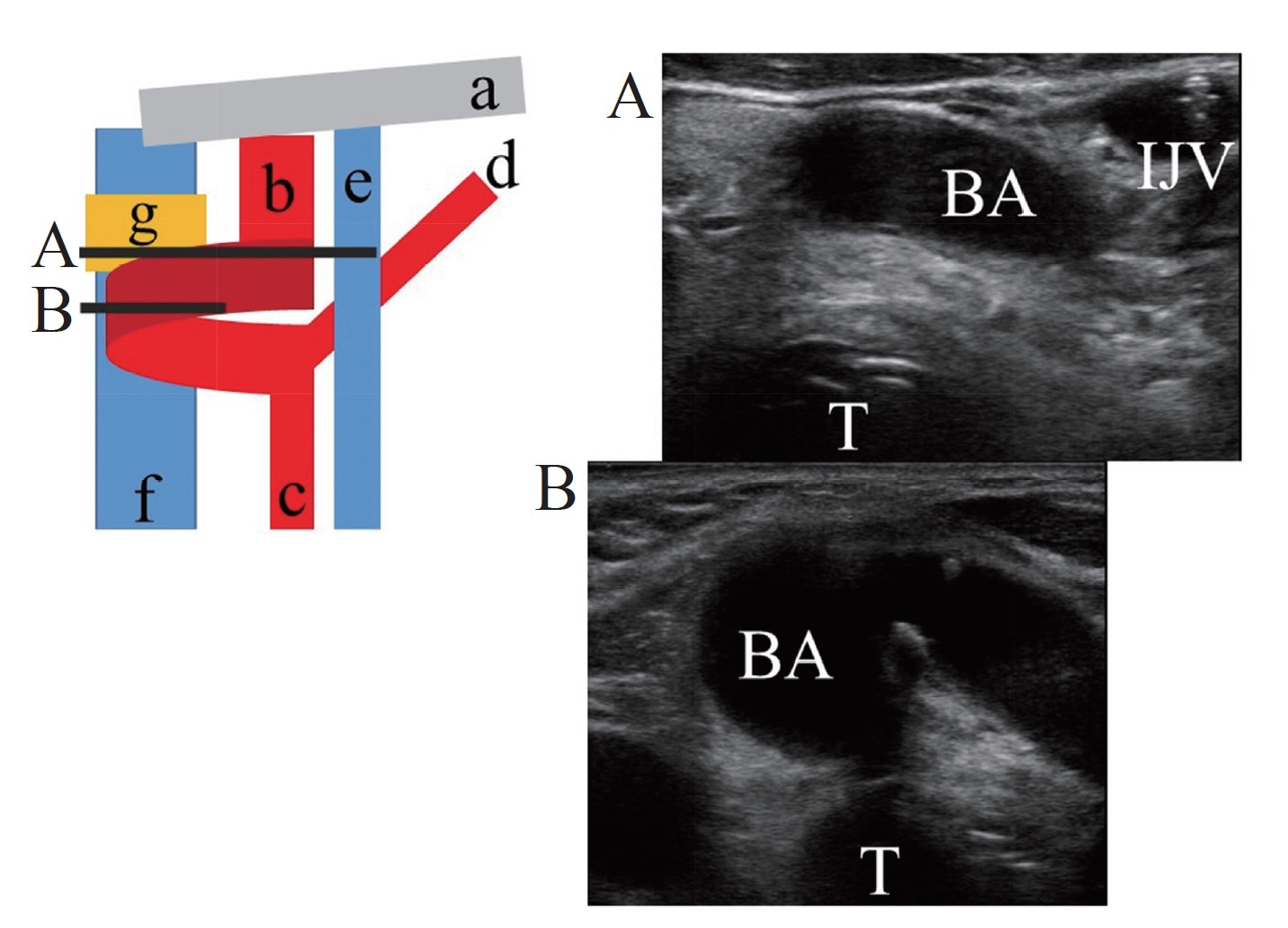
A 68-year-old woman underwent ultrasonography for determination of the spatial relationship between her right common carotid artery (CA) and right internal jugular vein for guidance in inserting the central venous catheter before surgery. The image showed that the brachiocephalic artery (BA) deviated to the midline from the cephalic side of the right clavicle (Fig. 1A). The BA coiled in front of the thyroid gland at the level of the 6th cervical vertebra (Fig. 1B), and diverged into the right common CA and right subclavian artery at the normal CA position.
Download full-size image
a: right clavicle; b: brachiocephalic artery (BA); c: common carotid artery; d: subclavian artery; e: internal jugular vein (IJV); f: trachea (T); g: thyroid gland.
Various anatomical variations of the BA have been described before. These include abnormal length, variations in position of the bifurcation point, atypical configurations, and variable size; but it is difficult to identify the frequency of each occurrence. A few previous reports showed that the carotid sheath unites with the visceral fascia at the level of the thyroid gland.1-3 During anesthesia, abnormal bifurcation and running direction of BA are rarely identified by pre-scanning ultrasonography prior to central venous catheterization. Therefore, the procedure to confirm the perivascular structures and spatial positions of both the common CA and the internal jugular vein is very important to avoid inadvertent puncture, catastrophic hemorrhage, and other complications.
Conflicts of Interest
Author declares no conflicts of interest.
References
| 1 |
Subasi ID, Yoruk O, Sipal S, Karakaya AD, Sengoz F.
Brachiocephalic artery anomaly at the neck: importance during minimally invasive video-assisted parathyroidectomy.
J Craniofac Surg 2013;24:e750–e773.
|
| 2 |
Hayashi S.
Histology of the human carotid sheath revisited.
Okajimas Folia Anat Jpn 2007;84:49–60.
|
| 3 |
Iwanaga J, Watanabe K, Tsuyoshi S, Tabira Y, Yamaki KI.
Tortuous common carotid artery: a report of four cases observed in cadaveric dissections.
Case Rep Otolaryngol 2016;2016:2028402.
|